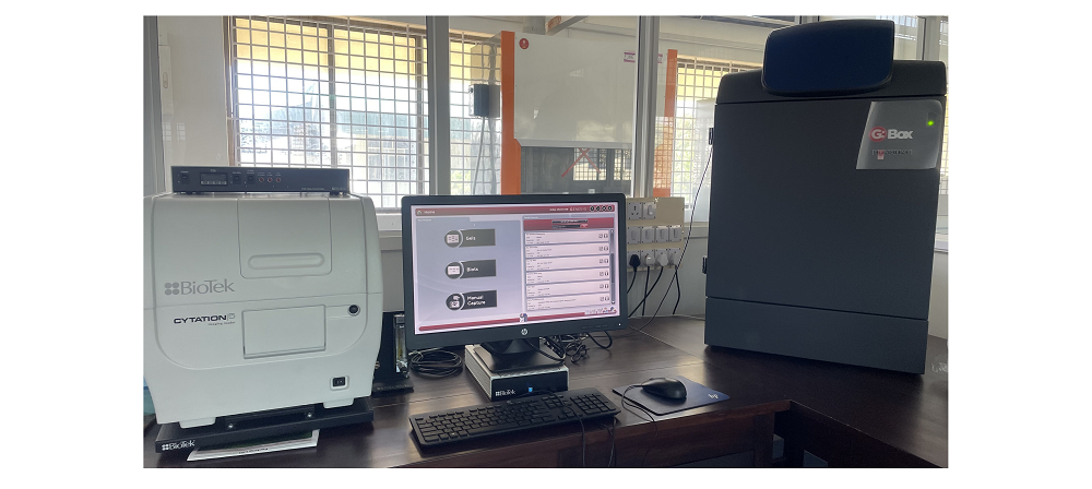
Cell Imaging Multi-Mode Reader
[CYT5MPV, BioTek Instruments, USA]
Located at: CRF Annexe - S01
I-STEM Equipment Code : 2938918 (This code can be used to locate the equipment on the I-STEM portal)
Facilities:
Module supports detection modes: UV-Vis absorbance, Fluorescence Luminescence, Time-resolved fluorescence
Read methods: Endpoint, kinetic, spectral scanning, well area scanning.
Applications:
• Absorbance, fluorescence and luminescence-based endpoint and kinetic assays
• Cell imaging: 6 to 1536 well plates
• Determination of cell count
• Cytoplasm, intracellular, subpopulation analysis,
• Signal translocation
• Cell migration and invasion
• Immunofluorescence
• Phenotypic assays
• Histology
Technical specifications Cell imaging multimode reader
• Detection mode: Fluorescence, Luminescence, UV-Vis absorbance and Time-resolved fluorescence
• Light sources: Xenon flash
• Detector: PMT for florescence system, Photodiode for absorbance
• Wavelength range: 250-700nm (fluorescence), 230-999nm (absorbance)
• Dynamic range: 7 decades (fluorescence), 0.4 OD (absorbance), >6 decades (luminescence)
• Reading speed (kinetic): 96 wells in 11 seconds, 384 wells in 22 seconds
• Assay type: End point, kinetic, spectral and well scanning read
• Plate compatibility: Reads 6 to 384 well plates and imaging of 6 to 1536 well plates
• It can accommodate microscope slides, petri and cell culture dishes, cell culture flasks (T25)
• Plate shaking: Linear, orbital, double orbital shaking modes
• Imaging modes: Fluorescence, bright-field, color bright-field and phase contrast with single color, multi-color, montage, time lapse and z-stacking
• Image filter cube: DAPI, GFP, Texas Red Filter cubes with imaging LED cubes of 365nm, 465nm and 590nm
• Monochromator wavelength accuracy and repeatability:2 nm, +0.2 nm
• OD accuracy: <1% at 2.0 OD <3% at 3.0 OD, OD linearity; <1% from 0 to 3.0 OD; OD repeatability:<0.5% at 2.0 OD
Testing charges.
|
Plate reading assay [per sample/plate] |
Cell imaging [per sample per mode] |
| NITK |
Rs. 300/- |
Rs. 600/- |
| Academic Institute* |
Rs. 660/- |
Rs. 960/- |
| R&D Institute* |
Rs. 780/- |
Rs. 1200/- |
| Industry* |
Rs. 900/- |
Rs. 1440/- |
*Sample preparation charges, GST(18%) extra. Above charges are for providing raw data.
For further Query Contact:
Ms. Jayashree Arun (Technical Staff, CRF), Email: crf_cellimg@nitk.edu.in
Dr.(Ms) Gangamma S (Cell-Imaging Faculty in-charge), Email: gangamma@nitk.edu.in, Mob: +91-8970110470
Prof. Keyur Raval, Professor InCharge-CRF, Email: keyurnraval@nitk.edu.in, Mob: +91-9342421444